Publications and Preprints

Olafson, E. R. , Georgia Russello, Jamison, K. W., Liu, H., Wang, D., Bruss, J. E., Boes, A. D., & Kuceyeski, A. (2021). Increased prevalence of a frontoparietal brain state at rest is associated with better motor recovery in individuals with pontine stroke affecting dominant-hand corticospinal tract. bioRxiv.
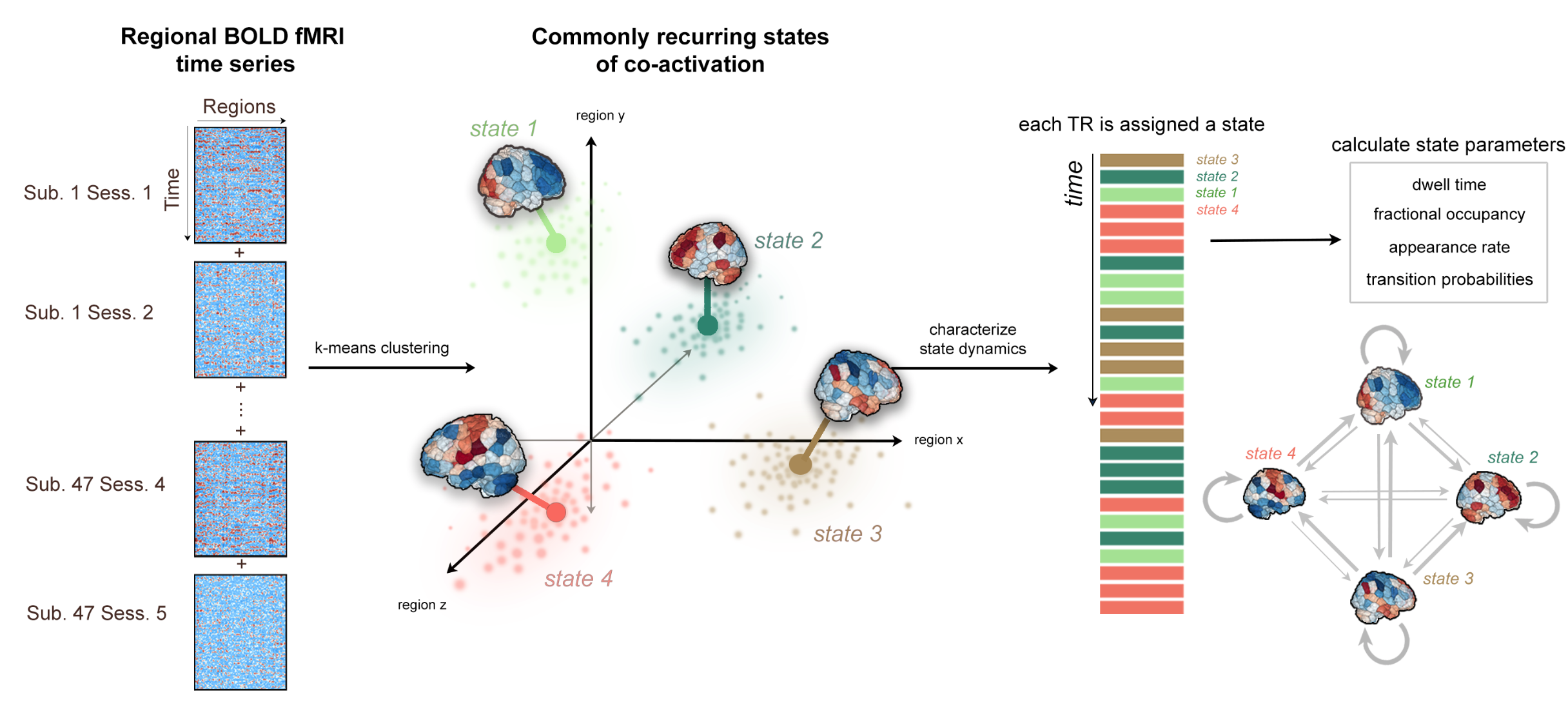
Although modulation of cortical activity is central to stimulation-based rehabilitative therapies, aberrant and adaptive patterns of brain activity after stroke have not yet been fully characterized. Here, we apply a brain dynamics analysis approach to study longitudinal brain activity patterns in individuals with ischemic pontine stroke. We first found 4 commonly occurring brain states largely characterized by high amplitude activations in the visual, frontoparietal, default mode, and motor networks. For individuals with dominant-hand CST damage, increased time spent in the frontoparietal state from 1 week to 3-6 months post-stroke was associated with better motor recovery over the same time period. This work suggests that when the dominant-hand CST is compromised in stroke, resting state configurations may shift to include increased activation of the frontoparietal network, which may facilitate compensatory neural pathways that support recovery of motor function when traditional motor circuits of the dominant-hemisphere CST are compromised

Olafson, E. R. , Jamison, K. W., Sweeney, E. M., Liu, H., Wang, D., Bruss, J. E., Boes, A. D., & Kuceyeski, A. (2021). Functional connectome reorganization relates to post-stroke motor recovery and structural disruption. Neuroimage.
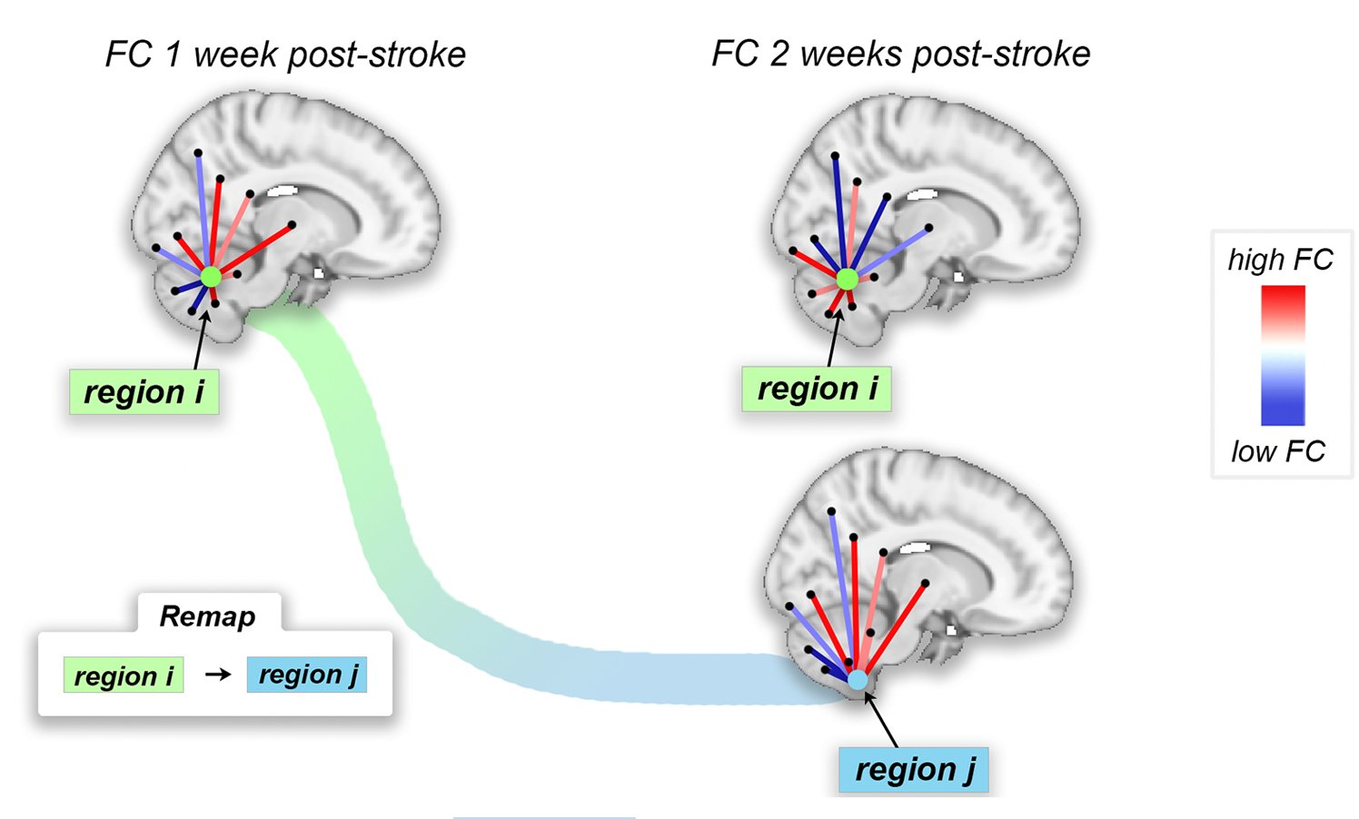
Remapping after stroke has not yet been observed with human fMRI. Here, we propose a novel biomarker for functional remapping using graph matching and apply it to a cohort of pontine stroke subjects. We found that, in general, remapping occurred in brain areas that were structurally damaged by the stroke, and that more remapping over time was correlated with better motor outcomes.
Undergraduate Work

Olafson, E. , Bedford, S. A., Devenyi, G. A., Patel, R., Tullo, S., Park, M. T. M., Parent, O., Anagnostou, E., Baron-Cohen, S., Bullmore, E. T., Chura, L. R., Craig, M. C., Ecker, C., Floris, D. L., Holt, R. J., Lenroot, R., Lerch, J. P., Lombardo, M. V., Murphy, D. G. M., Raznahan A., Ruigrok A. N. V., Spencer M. D., Suckling J., Taylor M. J., Lai M. C., Chakravarty, M. M. (2021). Examining the Boundary Sharpness Coefficient as an Index of Cortical Microstructure in Autism Spectrum Disorder. Cerebral Cortex, 31(7), 3338–3352.
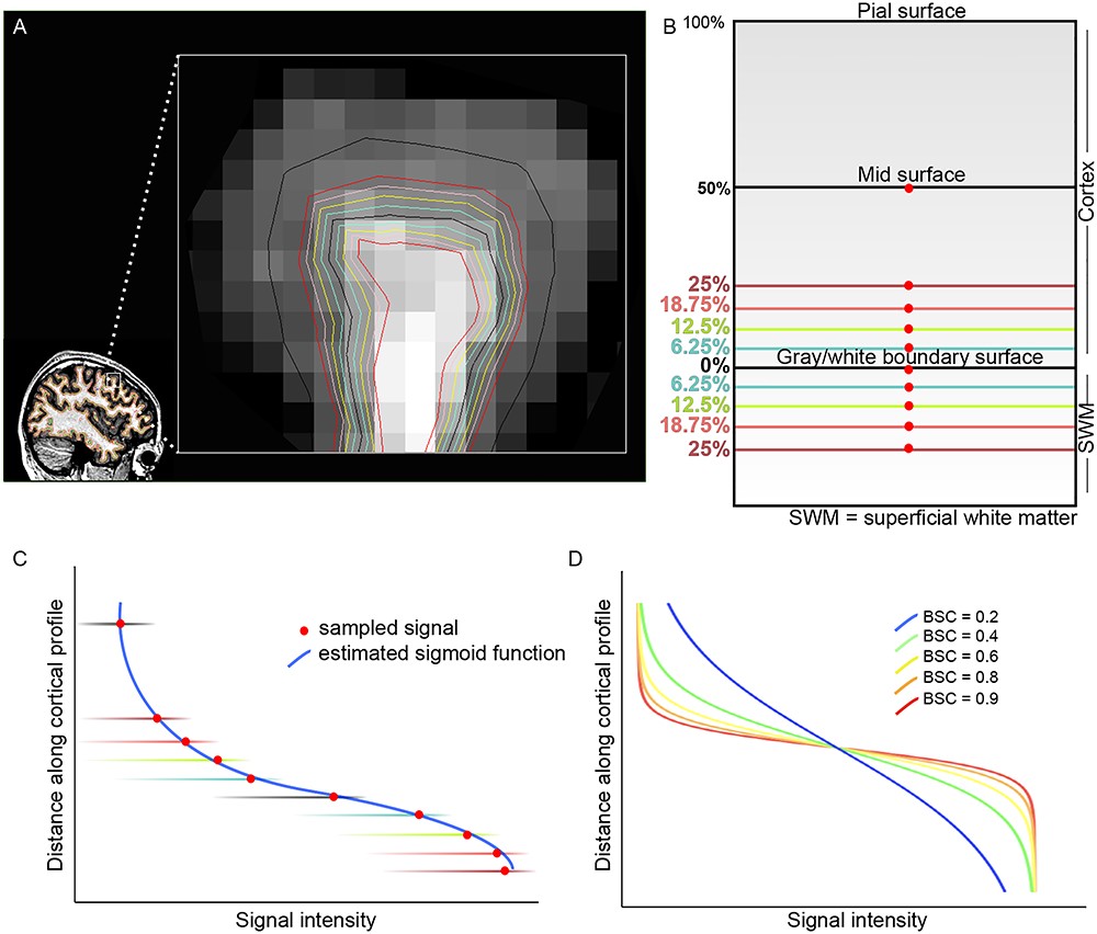
The phenotype of regionally specific increased cortical thickness observed in autism spectrum disorder (ASD) may be driven by several independent biological processes that influence the gray/white matter boundary. Here, we proposed to use the boundary sharpness coefficient (BSC), a proxy for alterations in microstructure at the cortical gray/white matter boundary, to investigate brain differences in individuals with ASD. We observed that individuals with ASD had significantly greater BSC indicating an abrupt transition (high contrast) between white matter and cortical intensities. Importantly, there was a significant spatial overlap between maps of the effect of diagnosis on BSC when compared with cortical thickness. These results invite studies to use BSC as a possible new measure of cortical development in ASD and to further examine the microstructural underpinnings of BSC-related differences and their impact on measures of cortical morphology.
Drakulich, S., Thiffault, A.-C., Olafson, E., Parent, O., Labbe, A., Albaugh, M. D., Khundrakpam, B., Ducharme, S., Evans, A., Chakravarty, M. M., & Karama, S. (2021). Maturational trajectories of pericortical contrast in typical brain development. NeuroImage, 235, 117974.